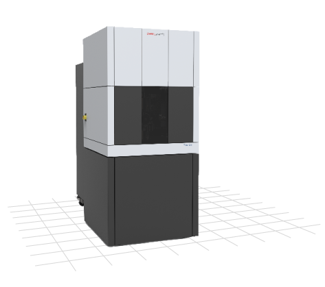
Talos™ F200i
The Thermo Scientific™ Talos F200i TEM is a 20–200 kV transmission electron microscope with a field electron gun (FEG).
The ThermoFisher Scientific™ Talos F200i TEM is a 20–200 kV transmission electron microscope with an FEG source, which allows high-quality STEM/TEM imaging with the Velox Software. Unique EDS absorption correction in Velox Software enables accurate quantification, while Thermo Scientific Maps Software for TEM and EDS provides intuitive image-based navigation over the entire sample. The system is equipped with the 4k×4k Ceta 16M camera. Single tilt and double tilt holders can be used for specimens analysis. The system features a Lorentz lens, allowing specimens to be observed in field-free conditions. This, in combination with electron holography, is a powerful tool for the visualization and analysis of magnetic domains.
Techniques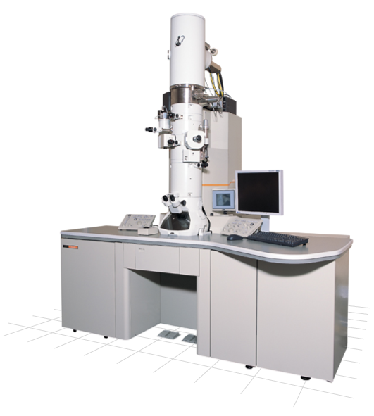
JEOL JEM-2100F
JEOL JEM-2100F TEM is a multipurpose analytical electron microscope with a field electron gun (FEG).
JEOL JEM-2100F TEM is a multipurpose analytical electron microscope with field electron gun (FEG) producing a highly stable and bright electron probe, essential for ultrahigh resolution in scanning transmission microscopy and in the analysis of nano-scaled samples. The microscope is equipped with an EDS detector for X-ray microanalysis with the Aztec analytical system, HAADF and BF detectors for STEM, an 8 Mpix camera by Gatan, and Digital Micrograph software. Single tilt and double tilt holders can be used for specimens analysis.
Techniques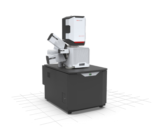
Apreo ChemiSEM
Scanning electron microscope with an FEG electron source, live elemental imaging, EBSD, and in-situ heating & tensile stages.
The ThermoFisher Scientific™ ChemiSEM system is equipped with in-beam and chamber detectors. The hybrid pixelated TruePix Direct Electron Detection EBSD is a high-speed, high-sensitivity direct electron detection system that provides comprehensive EBSD acquisition and data processing. The TrueSight LX EDS detector with ChemiPhase enables accurate identification of material phases with pixel-perfect precision, offering live acquisition via a novel data segmentation approach. The system also offers the possibility to heat (high-vacuum heating stage, up to 1000 °C) or mechanically pull (tensile module with narrow spindle distance 38 mm, EBSD-capable, capacity up to 5 kN) samples and perform in-situ experiments.
Techniques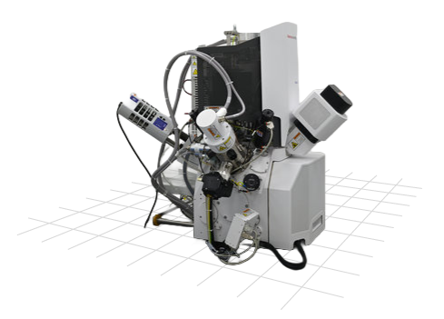
Scios 2 DualBeam
The Thermo Scientific™ Scios 2 DualBeam is an ultra-high-resolution analytical focused ion beam scanning electron microscopy (FIB-SEM) system.
The Thermo Scientific™ scanning electron microscope Scios 2 DualBeam is equipped with in-beam and chamber detectors. The system features a FIB which, among other things, allows — in combination with a gas injection system (GIS, deposition of C and W) and a nanomanipulator — material milling, 3D FIB-SEM tomography, and almost automatic preparation of TEM lamellas. The segmented BSE chamber detector is suitable for maximum materials contrast; an EDS detector with AZtec software provides chemical analysis; and the STEM detector allows imaging of the internal structure of TEM samples or nanostructures.
Techniques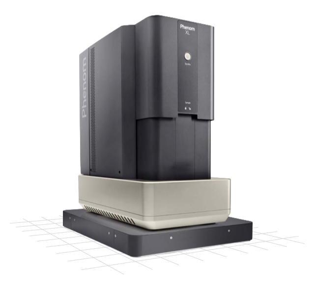
Phenom XL
Desktop SEM with a long-lifetime CeB6 source, fully integrated EDS, and large-sample capability up to 100×100 mm.
The ThermoFisher Scientific Phenom XL Desktop SEM system, with a long-lifetime thermionic source (CeB6), offers an SE detector for surface-sensitive imaging, a four-segment BSE detector that yields sharp images and provides chemical contrast information, and a fully integrated EDS system for elemental analysis. ChemiSEM software generates real-time, color-coded elemental maps directly over the SEM live image; combining SEM and EDS, it delivers ultra-fast, dynamically integrated results. The system also provides Maps 3 software for correlative electron microscopy and cross-platform imaging automation. The microscope can analyze large samples up to 100×100 mm at a resolution of 10 nanometres.
Techniques
Tescan LYRA 3 XMU
Scanning electron microscope Tescan LYRA 3 XMU FEG/SEMxFIB with an FEG electron source.
Scanning electron microscope Tescan LYRA 3 XMU FEG/SEMxFIB is equipped with EBSD Symmetry for crystallographic analysis and EDS UltimMax detectors with AZtec software for chemical analysis. The microscope also offers a FIB, allowing imaging and milling of the material by high-energy gallium ions. The FIB, in combination with a micromanipulator and gas injection system (GIS, deposition of Pt and W), can also be adopted for TEM sample preparation.
Techniques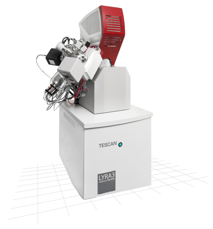
Tescan LYRA 3 XMH
Besides chamber SE and BSE detectors, the LYRA 3 XMH FEG/SEMxFIB is also equipped with in-beam SE, in-beam BSE, and STEM detectors.
The microscope offers a FIB, allowing imaging and milling of the material by high-energy gallium ions. The FIB, in combination with a micromanipulator and gas injection system (GIS, deposition of Pt and W), can also be adopted for TEM sample preparation. The microscope is equipped with an EDS X-Max detector and AZtec software for analysis and quantification of chemical composition. The AFM system NenoVision LiteScope can be placed into the microscope chamber, allowing for the simultaneous acquisition of AFM and SEM signals.
Techniques
SPM LiteScope
A Scanning Probe Microscope (SPM) designed for easy integration into scanning electron microscopes.
The combination of complementary AFM and SEM techniques makes it possible to use the advantages of both commonly used microscopy techniques. LiteScope incorporates a unique imaging technique — Correlative Probe and Electron Microscopy (CPEM) — enabling simultaneous acquisition of AFM and SEM data. LiteScope and the CPEM technology allow sample analysis in a way that was previously difficult or impossible with the two imaging technologies simultaneously.
- Surface topography measurements using AFM, incl. automated mapping of large surfaces
- Scanning tunneling microscopy (STM)
- Electrical surface characterization by measuring the EBIC current (see also MightyEBIC 2.0)
- Magnetic surface characterization using MFM
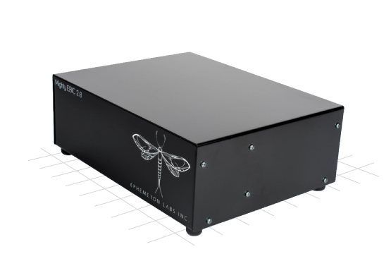
MightyEBIC 2.0
A scan controller and data acquisition interface for quantitative Electron Beam Induced Current (EBIC) measurements.
At the highest gain setting, this equipment achieves a current resolution of 0.76 fA. Using 18-bit AD converters provides over 64× higher sensitivity than most other scan controllers and a much wider dynamic range of 108 dB at each gain setting of the pre-amplifier. With 8 imaging inputs that sample simultaneously with a ±10 V range, the system can collect signals from secondary electrons (SE), backscattered electrons (BSE), and EBIC signals that are directly correlated in position. The system can interface directly with lock-in amplifiers to capture both amplitude and phase components. Two auxiliary voltage outputs with a ±10 V range allow biasing, backgating, or current–voltage sweeps to a device.
Techniques